RETURN TO TOP

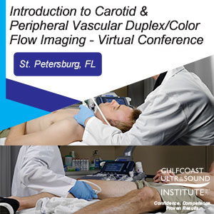
Introduction to Carotid & Peripheral Vascular Duplex/Color Flow Imaging - Virtual Conference

Introduction to Carotid & Peripheral Vascular Duplex/Color Flow Imaging

Dates: September 27 - 30, 2021


Introduction to Carotid & Peripheral Vascular Duplex/Color Flow Imaging

Please call 727-363-4500
To purchase this product


Introduction to Carotid & Peripheral Vascular Duplex/Color Flow Imaging

Dates: September 27 - 30, 2021

  • COMBINE COURSES & $AVE: When you Combine Back to Back Courses, Individual Course Tuition Receives a Discount!
  • Trusted Vascular Ultrasound CME Leader Since 1985
  • Most Comprehensive Vascular Ultrasound Courses Available
  • Master Level Vascular Ultrasound Faculty
  • Lifetime Access to E-Course Materials
  • Immediately Implement Skills Learned into Clinical Practice
  • Expand Diagnostic & Interventional Services Offered
  • ACCME Accredited with Commendation

Overview

Introduction to Carotid & Peripheral Vascular Duplex/Color Flow Imaging Virtual Conference is taught by leading vascular ultrasound experts, and is specifically designed for physicians, sonographers and other medical professionals who need Carotid & Peripheral Vascular Duplex/Color Flow Imaging training. These four day Carotid & Peripheral Vascular Duplex/Color Flow Imaging ultrasound courses includes 130+ interactive case presentations. In this (4) four day long vascular program, participants will learn ultrasound techniques for the entire vascular system.

Code: CP-211VC
ID: 5281

Schedule

      Download Course Itinerary

Topics

  • Doppler & Color Fundamentals
  • Carotid Anatomy & Physiology
  • Normal Spectral Analysis
  • Carotid Scan Protocol
  • Disease Progression & Stenosis Assessment
  • Carotid Imaging Pitfalls
  • Interactive Case Studies
  • Evaluation & Measurement of Intima-Media Thickness
  • Carotid Abnormalities
  • How to Structure a Report
  • Venous Anatomy, Scan Techniques & Normal Characteristics
  • Duplex / Color Evaluation of LE, UE DVT
  • Evaluation of Venous Insufficiency
  • Duplex / Color Evaluation for Venous Ablation Procedures
  • Types of Vein Ablation Procedures
  • Pre-Procedure Mapping
  • Post-procedure Mapping
  • LE Arterial Anatomy & Physiology
  • Clinical Exam & Indirect Testing
  • Direct Testing & Duplex Scanning
  • Live Demo: Arterial Duplex of Lower Arterial Disease

Objectives

  • Increase the participant’s knowledge to better perform and/or interpret Carotid Duplex/Color Flow Imaging ultrasound examinations.
  • Apply knowledge of the anatomy/physiology of the cerebrovascular system into the Carotid Duplex examination.
  • Cite Doppler/color physics principles and be able to (sonographers) apply these principles to optimize system controls or (physicians) utilize this information for identifying technical errors which may result in misdiagnosis.
  • Perform routine scan protocols and Doppler calculations in a complete carotid duplex/color examination.
  • Differentiate normal/abnormal spectral Doppler/color characteristics.
  • List methods for obtaining quantitative information and state the diagnostic relevance of each measurement.
  • Characterize plaque morphology and other pathology associated with cerebral vascular disease.
  • Perform Intima-Media Thickness measurements and state the clinical significance as a screening method for cardiovascular disease.
  • Integrate the information to include and prepare a structured report for a carotid ultrasound examination.
  • Apply diagnostic criteria for accurate interpretation of carotid duplex/color flow examinations.
  • Increase the participant’s knowledge to better perform and/or interpret upper and lower Peripheral Vascular ultrasound examinations.
  • Apply knowledge of the anatomy/physiology of the upper and lower extremity venous and arterial systems into the venous and arterial duplex and physiologic testing examinations.
  • Cite Doppler/color physics and be able to (sonographers) apply these principles to optimize system controls and/or (physicians) utilize this information for recognizing technical errors which may result in misdiagnosis.
  • Perform routine scan protocols, and document Doppler waveforms for lower extremity arterial and venous evaluations of the upper and lower extremity.
  • Differentiate normal/abnormal imaging, spectral Doppler and color characteristics for identifying arterial and venous disease.
  • State the indications and applications of indirect testing methods for lower arterial disease.
  • Demonstrate vein mapping techniques to identify suitability as a potential arterial bypass graft.
  • Perform routine scan protocols and document Doppler waveforms for venous evaluation of the lower extremities, including pre and post vein ablation evaluation.
  • State the role of ultrasound in the diagnosis and treatment of venous insufficiency.
  • Perform evaluation for venous insufficiency and patency of perforators for vein therapy treatment.

Tuition

Registration: $1,715.00

Audience

Sonographers, physicians, physician assistants and other medical professionals learning how to perform and/or interpret carotid and peripheral vascular duplex/color examinations. Participants may include (but are not limited to) those involved with internal medicine, cardiology, radiology, neurology, general/vascular surgery, and primary care.

Faculty

GulfCoast Ultrasound Institute

Phil Bendick  PhD, RVT, FSDMS, FSVU

Vascular Consultant.
Vass, NC.

No relevant financial relationships to disclose.

GulfCoast Ultrasound Institute

Lori Green  BA, RDMS, RDCS, RVT

President, Program Director.
Gulfcoast Ultrasound Institute, Inc.
Saint Petersburg, FL.
No relevant financial relationships to disclose.
GulfCoast Ultrasound Institute

Trisha Reo  AAS, RDMS, RVT

Program Coordinator
Gulfcoast Ultrasound Institute, Inc.
Saint Petersburg, FL.
No relevant financial relationships to disclose.

Faculty Disclosure
Gulfcoast Ultrasound Institute, Inc.adheres to ACCME Essential Areas, Standards, and Policies regarding industry support of CME. Speakers will disclose any relevant commercial relationships prior to the start of the educational activity.

Content Validation
All presentations for this CME activity were reviewed and approved by members of the GUI CME Review Committee to determine content validity and to ensure that no conflicts of interest exist prior to the activity commencement or material compilation.

Disclosure of Individuals in Control of Content
In addition to the faculty listed the following individuals are recognized by GUI as being in control of content of this program:

James Mateer, MD, RDMS (Medical Director-planner, QI Task Force Subcommittee)
Medical Director, Gulfcoast Ultrasound Institute
Milwaukee, WI
No relevant financial relationships to disclose.
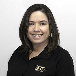
Charlotte Derr, MD, RDMS, FACEP, FPD-AEMUS (Co-Medical Director-planner & QI Task Force Subcommittee)
Associate Professor of Emergency Medicine
Fellowship Director of Advanced Emergency Medicine Ultrasound Fellowship Program
University of South Florida Morsani College of Medicine
Tampa, FL
No relevant financial relationships to disclose

Andreas Dewitz, MD, RDMS (Member of Advisory Board & QI Task Force Subcommittee)
Associate Professor of Emergency Medicine
Vice Chair of Ultrasound Education
Boston Medical Center, Boston, MA.
No relevant financial relationships to disclose

Lori Green, BA, RDMS, RDCS, RVT (Program Director - planner, Content Reviewer, QI Task Force Subcommittee)
Program Director and President,
Gulfcoast Ultrasound Institute, Inc.
St Petersburg, FL.
No relevant financial relationships to disclose

Trisha Reo, AAS, RDMS, RVT (Program Coordinator - planner, Content Reviewer, QI Task Force Subcommittee)
Gulfcoast Ultrasound Institute, Inc.
St Petersburg, FL.
No relevant financial relationships to disclose


Disclosure of Commercial Support
There was no commercial support involved in the planning, development/implementation of this activity.


Credits

The Gulfcoast Ultrasound Institute is accredited by the Accreditation Council for Continuing Medical Education (ACCME) to provide continuing medical education for physicians.

Introduction to Carotid Duplex/Color Flow Ultrasound

The Gulfcoast Ultrasound Institute is accredited by the Accreditation Council for Continuing Medical Education (ACCME) to provide continuing medical education for physicians.

The Gulfcoast Ultrasound Institute designates this internet live activity for a maximum of 8.00 AMA PRA Category 1 Credit(s)™. Physicians should claim only the credit commensurate with the extent of their participation in the activity.

This course also meets CME / CEU requirements for ARDMS.Note: While offering the CME credit hours noted above, activities are not intended to provide extensive training or certification for exam performance or interpretation.

Introduction to Peripheral Vascular Ultrasound

The Gulfcoast Ultrasound Institute is accredited by the Accreditation Council for Continuing Medical Education (ACCME) to provide continuing medical education for physicians.

The Gulfcoast Ultrasound Institute designates this internet live activity for a maximum of 8.00 AMA PRA Category 1 Credit(s)™. Physicians should claim only the credit commensurate with the extent of their participation in the activity.

This course also meets CME / CEU requirements for ARDMS.Note: While offering the CME credit hours noted above, activities are not intended to provide extensive training or certification for exam performance or interpretation.

Doppler and Color Fundamentals: Vascular Ultrasound - Online Video

The Gulfcoast Ultrasound Institute is accredited by the Accreditation Council for Continuing Medical Education (ACCME) to provide continuing medical education for physicians.

The Gulfcoast Ultrasound Institute designates this internet activity enduring material for a maximum of 2.00 AMA PRA Category 1 Credit(s)™. Physicians should claim only the credit commensurate with the extent of their participation in the activity.

This course also meets CME / CEU requirements for ARDMS.Note: While offering the CME credit hours noted above, activities are not intended to provide extensive training or certification for exam performance or interpretation.

Resources

39 YEARS

Serving the Medical Community

180,000+

Participants Trained

500,000+

CME Credits Awarded

6,000+

Courses Offered

CONFIDENCE, COMPETENCE, PROVEN RESULTS

(727) 353-8222 - Google Ads