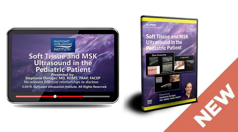
Soft-Tissue and MSK Sonography in the Pediatric Patient Training Video is designed to present ultrasound applications for evaluation of soft-tissue and MSK pathology in the pediatric patient.
Objectives:
1. State indications and applications for ultrasound evaluation of soft tissue abnormalities.
2. List applications for the focused MSK ultrasound exam.
3. Identify ultrasound characteristics of cellulitis and abscess.
4. State the benefits of using ultrasound for foreign body localization and removal.
5. Recognize ultrasound findings associated with fracture assessment and use in reduction techniques.
6. State benefits of ultrasound-guided nerve blocks.
Topics:
• Cellulitis
• Necrotizing Fasciitis
• Abscess
• Foreign Body Localization/Removal
• Fracture Evaluation and Reduction
• Tendon Injuries
• Nerve Blocks
• Joint Effusion: Hip & Knee
Audience:
Physicians, PA’s, sonographers and other medical professionals who will be involved with performing and/or interpreting pediatric emergency/critical care ultrasound examinations. Physicians may include (but is not limited to) emergency medicine, critical care, trauma surgery, radiology, internal medicine, and primary care.
(727)363-4500
|
CREATE ACCOUNT
|
LOG IN
|
CART
|
Item(s) added to cart
Proceed to Checkout
Continue Shopping


