Let’s walk through a practical approach to using the subcostal window to help differentiate between normal SVC flow and a possible patent foramen ovale (PFO). This technique is straightforward but often overlooked, and it can make your imaging both clearer and more accurate.
Starting With the Basics
When scanning from the subcostal window, we typically begin by visualizing the superior vena cava (SVC) flow as it enters the right atrium. It’s standard practice to interrogate the interatrial septum closely, especially around the area known as the fossa ovalis, where an echo dropout is often seen. This area is clinically significant because it’s the common location for a PFO to occur.
Applying Color Flow Doppler
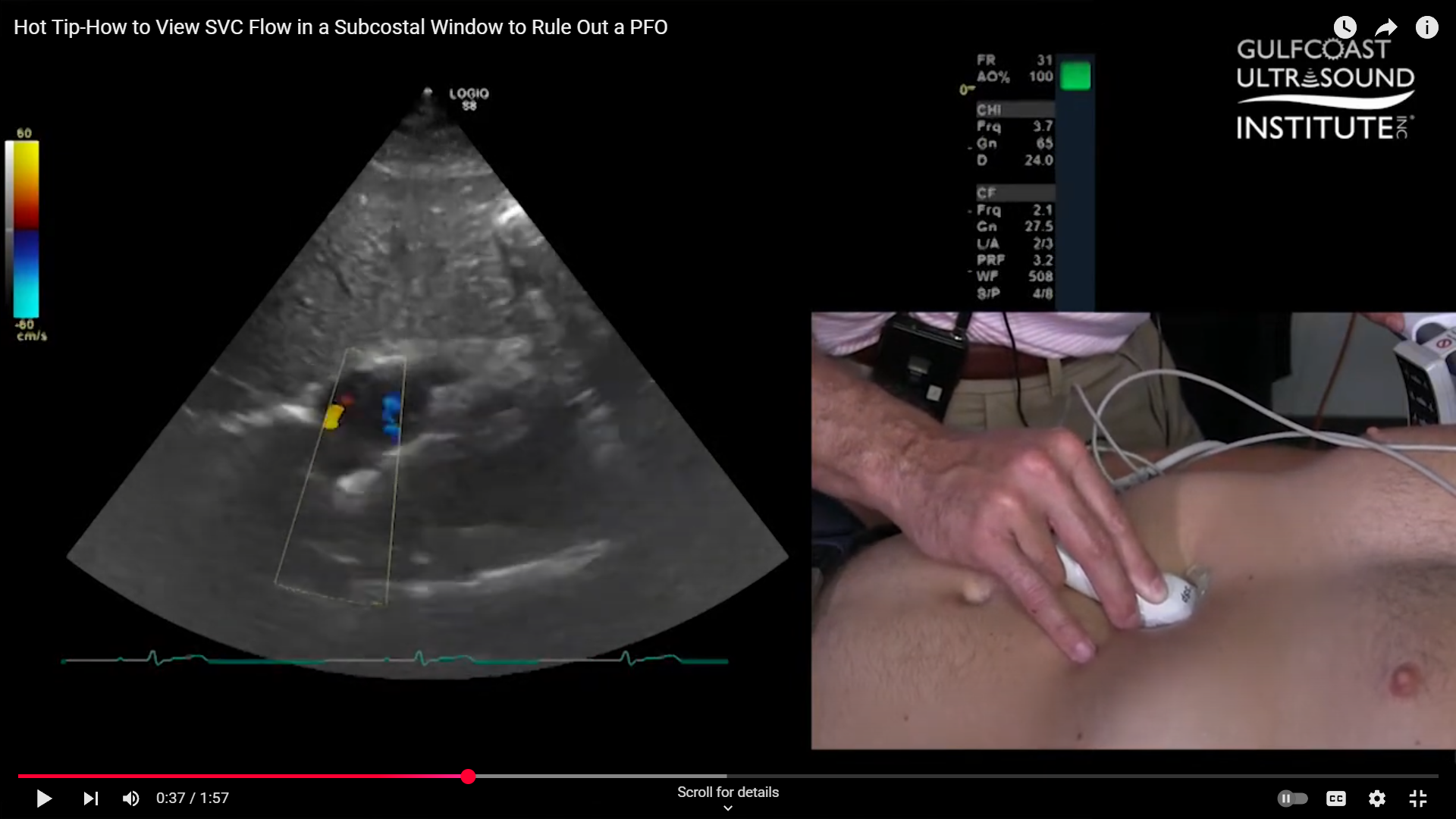
Once you’ve identified the SVC, applying color flow Doppler can help visualize the blood flow pattern. Normally, you’ll see red laminar flow moving into the right atrium. However, distinguishing between this normal SVC flow and a PFO shunt can be challenging. Sometimes, the color Doppler image isn’t clear enough to show whether there’s a true PFO or just normal flow dynamics.
The Key Adjustment for Better Imaging
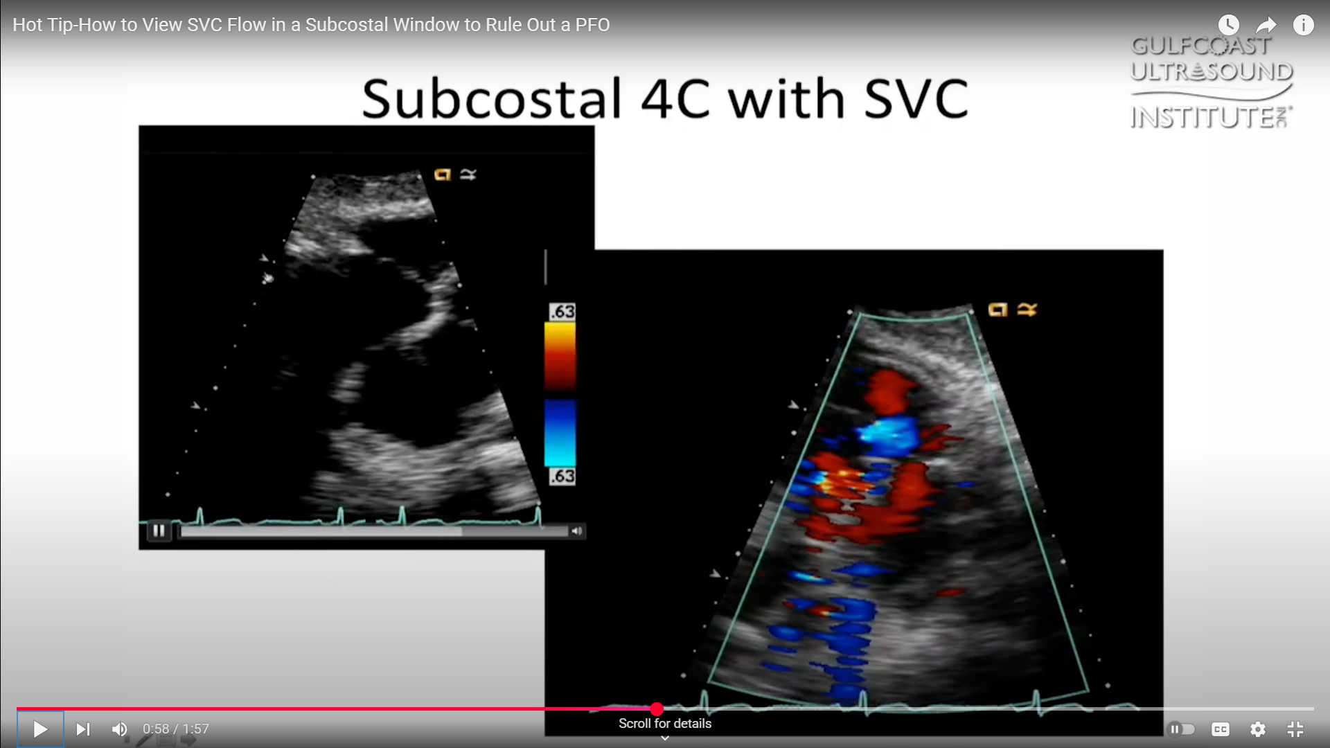
Here’s where a small adjustment can make a big difference. From your standard three o’clock view of the subcostal window, rotate your probe toward the five o’clock position. By making this shift, you bring the bicaval view into focus, allowing you to see both the inferior vena cava (IVC) and SVC together. On your screen, you’ll notice the SVC flow coming up and curving slightly to the right.
In this view, you’ll often be able to see the small PFO flow separate from the normal SVC flow. On a still frame, the SVC flow should appear as a consistent laminar pattern, while the PFO flow will be visible as a distinct jet at the level of the fossa ovalis. This adjustment provides a clearer way to differentiate between these two important findings.
Putting It Into Practice
Like any skill in ultrasound, it takes practice to become confident. Try rotating your probe during your next exam and see how the bicaval view sharpens your understanding of the anatomy and flow patterns. Over time, this small technique can help you rule out a PFO more reliably and improve the quality of your cardiac imaging.
Your Next Step in Ultrasound Mastery
Ready to sharpen your ultrasound skills further? Call the Gulfcoast Ultrasound Institute at Ph: 727-363-4500 for all of your ultrasound training needs! We’re conveniently located at 111 2nd Ave NE, #800, St. Petersburg, FL 33701.
Whether you’re just starting out or looking to deepen your expertise, we’re here to help you keep growing in your practice.