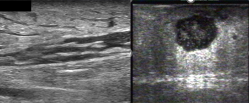
The use of Point-of-Care Ultrasound (POCUS) can be a useful tool for differentiating between abscesses and cellulitis, which are both common soft-tissue infections frequently seen in the emergency and family medicine care settings.
The diagnosis of cellulitis by physical exam alone is typically straight-forward when classic signs of erythema, warmth, and tenderness are present.
Findings during a physical exam can however be misleading in various situations such as a cutaneous abscess early in formation, small size, located in an area of thick subcutaneous fat or deep to the skin surface.
There are a variety of benefits for the use of POCUS for assessment of skin and soft-tissue infections (SSTI):
- Determine if an abscess is present
- Reduce time to initiate appropriate treatment for improved patient outcomes and safety
- Determine the best location for an incision to avoid penetrating blood vessels, nerves, and tendons
Scan Technique
Appropriate scan techniques and ultrasound system optimization is essential to obtain high-quality, diagnostic images for accurate diagnosis.
The exam should be performed with a high frequency linear array transducer for imaging superficial structures. In some cases, such as digits, a standoff gel pad or water-bath technique may be needed to effectively evaluate the region of interest. It is important to scan the area in longitudinal and transverse scan planes to accurately delineate the abscess/lesion.
Image Characteristics
It is important to be familiar with the ultrasound characteristics associated with normal skin and soft-tissue anatomy in order to recognize the appearance of pathology.
Normal Skin and Soft-Tissue Sonographic Characteristics
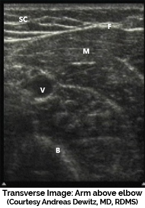
The skin appears as a thin, homogeneous, and hyperechoic line immediately under the transducer. The subcutaneous tissue (SC) is seen below the skin surface as hypoechoic fat and echogenic septae separating fat lobules. The fascia (F) appears as an echogenic line over the muscle surface. The muscle (M) is hypoechoic with internal hyperechoic (echogenic) striations. Blood vessels (V) appear anechoic (black with no internal echoes) and the bone (B) is hyperechoic with posterior shadowing.
Cellulitis
Example A
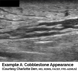
Cellulitis, appears as "cobblestone" on ultrasound, with hyperechoic subcutaneous tissue intersected by anechoic fluid. In addition, there is no well-defined fluid pocket. A similar pattern is seen in any edematous condition.
Example B
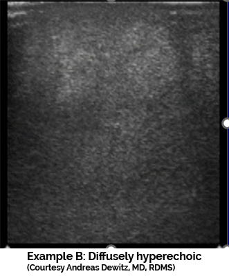
Cellulitis may also appear on the ultrasound exam as a diffusely hyperechoic soft tissues with poor detail resolution due to extensive tissue edema.
Abscess
On ultrasound, abscesses appear as spherical or oblong collections of fluid that are anechoic or hypoechoic, and may contain hyperechoic debris. Motion of the abscess cavity contents may be elicited with gentle probe pressure (Swirl Sign). The fluid can be uniform throughout, or it may have areas with internal echoes that make it heterogeneous. In addition posterior acoustic enhancement may be present.
Example A
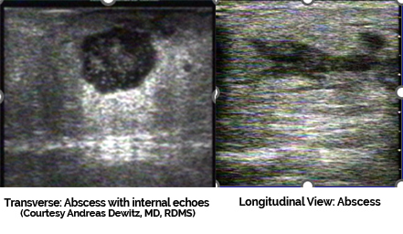
Example B
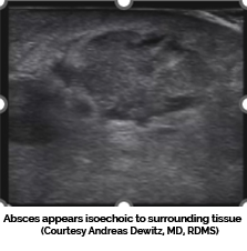
Abscess contents may on occasion appear isoechoic with surrounding tissue. Slight probe pressure will elicit the “swirl sign” to improve diagnostic accuracy.
Summary
POCUS evaluation of skin and soft-tissue infections is fast, cost-effective, portable, and readily available in many practice settings. Studies have revealed POCUS is effective to decrease time to treatment, avoid unnecessary procedures, and improve patient safety and satisfaction.
References:
O. Jon Ma, MD., James R. Mateer, MD. Emergency Ultrasound 4th ed. 2021. New York: McGraw Hill. 2021.
Want to obtain the skills needed to accurately differentiate skin, soft-tissue infections and more?
Attend a Gulfcoast Ultrasound Institute, Inc. (GCUS) hands-on skills training program. A variety of dates and education formats are available to best meet your needs at our education facility located in sunny St. Petersburg, Florida.
Upcoming GCUS Course dates
Introduction to Emergency Medicine Ultrasound
September 9-11, 2024: (Traditional Education Format)
November 14-15, 2024: (Blended Education Format)
May 1-2, 2025: (Blended Education Format)
Introduction and Advanced Emergency Medicine Ultrasound
November 14-15, 2024: (Blended Education Format)
February 10-14, 2025: (Traditional Education Format)
May 1-2, 2025: (Blended Education Format)
POCUS for Family and Internal Medicine
November 14-15, 2024: (Blended Education Format)
May 1-2, 2025: (Blended Education Format)
June 11-13, 2025: (Traditional Education Format)
Can’t Travel? We will come to you! Group training is available on a request basis.
Prefer one-on-one training? We offer Private one-on-one customized blended education format programs at the GCUS education facility.
Contact us today for more details!
