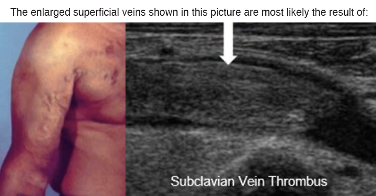
A. Central vein obstruction
B. Clotted dialysis access
C. Basilic vein thrombus
D. Prior thrombophlebitis
Answer at bottom of article.
Patients with upper extremity DVT will typically present with arm swelling and prominent superficial veins.
Paget-Schroetter syndrome, also known as effort thrombosis, refers to primary thrombosis of the axillary and/or subclavian vein. It can be thought of as a venous equivalent of thoracic outlet syndrome. Central venous catheters and compression of the subclavian vein when it passes through the costoclavicular space are the most favored mechanisms of thrombosis. Upper extremity DVT may also occur due to an underlying condition such as certain cancers, trauma, or radiation therapy.
Introduction to Peripheral Vascular Duplex/Color Flow Imaging
Introduction to Peripheral Vascular Duplex/Color Flow Imaging Live Training Course is taught by leading vascular ultrasound experts and includes topics pertaining to the AIUM, ACR, and IAC ultrasound practice and lab accreditation guidelines. Lectures, and interactive case studies pertaining to upper and lower extremity arterial and venous applications included. Extensive hands-on scanning is provided with the industry lowest 3:1 participant to faculty hands-on scan ratio. The program also includes a review of 65+ peripheral vascular ultrasound examinations to improve competence to perform and/or interpret peripheral vascular ultrasound examinations.
For the most comprehensive vascular ultrasound training, combine the Peripheral Vascular course with the 2-day Carotid Duplex/Color Flow Imaging program (total 4-day program) to expand your competence to increase patient services. The combined course offers a tuition discount and cost-saving with travel expenses.
- FREE Pre Course Online Video: Doppler & Color Fundamentals
- Destination Education in St. Petersburg, Florida
- Most Comprehensive Peripheral Vascular Ultrasound Course Available
- Trusted Vascular Ultrasound CME Leader Since 1985
- Industry Leading 3:1 Participant to Faculty Hands-On Scan Ratio
- Scan Groups Assigned Based on Specialty Practice and Level of Experience
- Lifetime Access to E-Course Materials
- ACCME Accredited with Commendation
- Master Level Vascular Ultrasound Faculty
- State-Of-The-Art Lecture Facility and Scan Labs
- Choose from Multiple Leading Manufacturer Ultrasound Equipment
- Immediately Implement Skills Learned into Clinical Practice
- Expand Diagnostic & Interventional Services Offered
- Live Demonstration: Venous Insufficiency
- Free CME-Z Membership with Registration (Save 5-10% off all future Gulfcoast product purchases)
Correct Answer: A


