(727)363-4500
|
CREATE ACCOUNT
|
LOG IN
|
CART
|
Item(s) added to cart
Proceed to Checkout
Continue Shopping
Ends in
(15% off Online Courses , Workbooks, Mock Exams, and Flashcards)
Discount applied at checkout

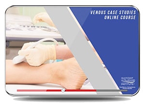
Venous Case Studies Online Course has been designed to provide a large variety of cases in order to improve competence in the diagnosis/interpretation of venous ultrasound pathologies. Recorded during a live Vascular Case Review program, our expert faculty presents 29 cases in a “double-read” format. The patient history/physical examination is provided along with the corresponding ultrasound case documentation.
Using the interactive format, participants will answer one or more multiple choice questions pertaining to each case. After submitting the answer to each question, the correct diagnosis will be discussed including correlation with other diagnostic modalities. The learner's responses to each question are recorded in order to provide a case log report of the exams interpreted. The “double read” case log may be used to meet hospital credentialing, individual certification*, and/or lab accreditation requirements.
Date of Original Release: 4/10/2019
Reviewed for content accuracy: 4/10/2022
Reviewed by: Gulfcoast Ultrasound CME Committee
Re-Reviewed for content accuracy: 4/10/2025
Re-Reviewed by: Gulfcoast Ultrasound CME Committee
This edition valid for credit through: 4/10/2026
Registration: $330.00
(12 Months Unlimited Access)

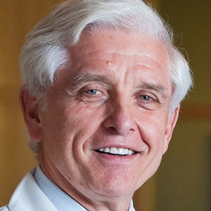
Dennis Bandyk MD, FACS
Professor of Surgery, University of San Diego.
Director of Vascular Surgery Division.
San Diego, CA.
No relevant financial relationships to disclose.


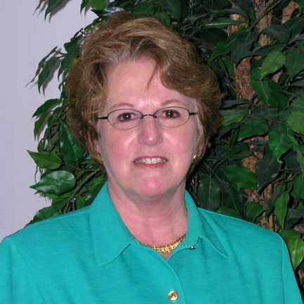
Marsha M. Neumyer BS, RVT, FSDMS, FSVU, FAIUM
Assistant Professor of Surgery at Penn State’s College of Medicine.
Former Director of the Vascular Lab Section of the Penn State Vascular Institute, Milton S. Hershey Medical Center.
International Director of Vascular Diagnostic Educational Services
Director and CEO of Vascular Resource Associates.
Past President of the Society of Vascular Technology and a founding member of the Intersocietal Commission for Accreditation of Vascular Laboratories (ICAVL)
Fellow of the Society of Vascular Technology
Fellow and member of the Board of Governors of the American Institute of Ultrasound in Medicine
1997 recipient of the Joan Baker Pioneer Award given by the Society of Diagnostic Medical Sonography
Ultrasound Consultant for Unetixs Vascular Inc.
Hershey, Pennsylvania.
GUI QI Task Force Subcommittee
No relevant financial relationships to disclose.


Robert Atnip MD, RVT, RPVI
Professor Emeritus, Department of Surgery and Radiology
Division of Vascular Surgery
The Milton S. Hershey Medical Center
The College of Medicine
The Pennsylvania State University
GUI QI Task Force Subcommittee
No relevant financial relationships to disclose.

James Mateer, MD, RDMS (Medical Director - planner, QI Task Force)
Gulfcoast Ultrasound Institute
Milwaukee, WI
No relevant financial relationships to disclose.
Charlotte Derr, MD, RDMS, FACEP (Co-Medical Director-planner, QI Task Force)
Assistant Professor of Emergency Medicine &
Fellowship Director of Emergency Medicine
Ultrasound Fellowship Program
University of South Florida Medical School
Tampa, FL
No relevant financial relationships to disclose
Andreas Dewitz, MD, RDMS(Member of Advisory Board, QI Task Force Subcommittee)
Associate Professor of Emergency Medicine
Vice Chair of Ultrasound Education
Boston Medical Center Boston, MA
No relevant financial relationships to disclose
Lori Green, BA, RT, RDMS, RDCS, RVT (Program Director-planner, Content Reviewer, QI Task Force)
President, Gulfcoast Ultrasound Institute, Inc.
No relevant financial relationships to disclose
The Gulfcoast Ultrasound Institute is accredited by the Accreditation Council for Continuing Medical Education (ACCME) to provide continuing medical education for physicians.
The Gulfcoast Ultrasound Institute designates this enduring material for a maximum of 5.00 AMA PRA Category 1 Credit(s)™. Physicians should claim only the credit commensurate with the extent of their participation in the activity.
This course also meets CME/CEU requirements for ARDMS. Note: While offering the CME credit hours noted above, activities are not intended to provide extensive training or certification for exam performance or interpretation.
Serving the Medical Community
Participants Trained
CME Credits Awarded
Courses Offered
(727) 353-8222 - Google Ads